Thank you for your question, Nicole, Ivanne, and Juan.
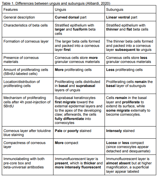
The differences in size and shape of the beta cells found in unguis and that of the subunguis can be explained by their differences in beta-cell proliferation and movement pattern (Alibardi, 2020). Unguis exhibits higher proliferation during development and growth and is composed of a compact corneous layer made of separated but tightly interlocked corneocytes. Conversely, subunguis displays lower proliferation and is characterized by a softer layer whose superficial corneocytes desquamate. Moreover, their movement pattern differs such that the proliferating beta-cells in the unguis are moving within epithelial layers, probably toward the apex of the developing claw. Meanwhile, beta-cells from the tip of the unguis move downward into the distal-most region of the subunguis (Alibardi, 2020).
Consequently, more beta cells in the unguis imply faster growth than the subunguis, allowing the former to be curved all around the end of the digit at the same time, covering the latter. Meanwhile, the thin corneous layer in the subunguis implies the initial presence of keratohyalin-like granules, which are storage vesicles for major corneocyte proteins (Alibardi, 2008; Amen et al., 2013). Notably, the combined movement of the beta-cells in the unguis and subunguis contributes to sharpening the crocodilian claw which is necessary to achieve its function, such as the grasping and tearing of a prey (Alibardi, 2020).
References:
Alibardi, L. (2008). Cornification in developing claws of the common Australian skink (Lampropholis guichenoti) (Squamata, Lacertidae). Italian Journal of Zoology, 75(4), 327–336. https://doi.org/10.1080/11250000801973334.
Alibardi, L. (2020). Immunolocalization of corneous beta proteins of the Epidermal Differentiation Complex in the developing claw of the alligator. Annals of anatomy, 231, 151513. https://doi.org/10.1016/j.aanat.2020.151513
Alibardi, L. (2021). Development, structure, and protein composition of reptilian claws and hypotheses of their evolution. Anatomical record (Hoboken, N.J. : 2007), 304(4), 732–757. https://doi.org/10.1002/ar.24515
Amen, N., Mathow, D., Rabionet, M., Sandhoff, R., Langbein, L., Gretz, N., Jäckel, C., Gröne, H. J., & Jennemann, R. (2013). Differentiation of epidermal keratinocytes is dependent on glucosylceramide:ceramide processing. Human molecular genetics, 22(20), 4164–4179. https://doi.org/10.1093/hmg/ddt264